Introduction:
Chromatography is a group of processes that are used in analytical chemistry to separate a mixture into its individual components. There are several types of chromatography the two most commonly used are probably gas chromatography (GC) and high performance liquid chromatography (HPLC). In general, chromatographic systems have four main components: a column or capillary, a stationary phase (non-moving) in the column or capillary, a mobile phase that flows through the column or capillary and a way to detect compounds at the end of the column or capillary. The basic principle of chromatography is that different compounds in a mixture spend different amounts of time in the stationary phase and therefore, take different amounts of time to reach the end of the column or capillary. The amount of time a compounds spends in the stationary phase depends on one or more chemical or physical properties of the compound including, but not limited to: size, molecular weight, polarity and shape.
GC is used to separate gases or liquids that can be heated to the vapor phase without decomposing. The mobile phase of GC is usually an inert gas such as helium and simply carries the compounds down the column or capillary when the compound is not adsorbed (stuck to) the stationary phase. The stationary phase of a GC separation can vary dramatically depending on the properties of the molecules to be separated. When a compound leaves the column or “elutes” it then passes through a detector that senses the presence of the compound and registers a response. There are many detectors that can be used for GC. A thermal conductivity detector (TCD) can be used to detect most molecules. Other detection methods are more specific to the compounds, for instance a flame ionization detector (FID) or a catalytic combustion detector (CCD) are great detectors for hydrocarbons (i.e. propane, butane, octane) but would not be able to detect other compounds such as H2 or N2O since they do not contain any carbon. A plot of the detector response versus time is called a chromatogram. The time that a compound elutes
Property Buck Scientific, Inc. East Norwalk, CT 06855 800-562-5566
(shows a peak on the chromatogram) from the column can be used to identify the compound for qualitative analyses. The area of each peak can be used to determine the amount of compound present in the original mixture (quantitative analysis) as long as a calibration curve is prepared first relating the peak area to the concentration of the analyte.
Purpose:
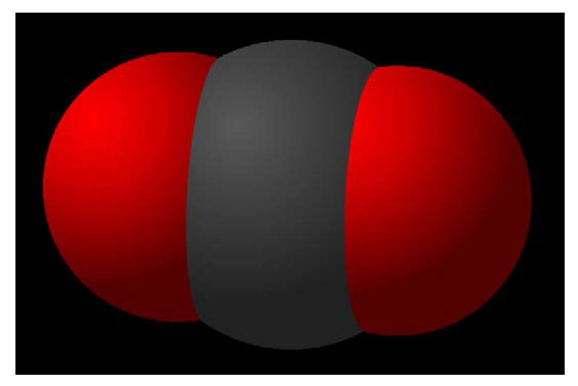
Carbon dioxide (CO2) is a chemical that has been in the news a considerable amount in recent years. This new exposure is due to its implication in global warming. The amount of carbon dioxide in air can be measured using GC. This proves useful in determining the amount of carbon dioxide in such environments as an open room, human breath, exhaust from several models and years of automobiles or even near trees. This experiment will allow for the determination of relative concentrations of CO2.
Instrumentation:
Buck Model 310/910 Gas Chromatograph (GC) with a thermal conductivity detector (TCD).
10' x 1/8" SS column packed with HayeSep D 100/120 mesh
Peak Simple Software
100 μl syringe
Samples:
Samples from various sources that will have carbon dioxide in them (some examples were given in the introduction, but use your imagination to think of places that may have varying amounts of CO2)
Safety:
Wear goggles at all times in the lab. Always follow good laboratory practices.
Procedure:
Instrument Setup:
Follow the specific instrument procedures in the manual for initial instrument set up
Turn on GC
Open Peak Simple Software and ensure proper setup of software interface (see Addendum below for instructions on how to properly set up Peak Simple)
Set the temperature to 80°C for 10 minutes. (Note: This may be altered to optimize the separation)
Standard Preparation:
If CO2 is available, you can prepare varying concentrations in carrier gas to construct a calibration curve in order to determine exact amount of carbon dioxide in the samples. However, the relative amount of CO2 can be measured without constructing a calibration curve and running standards by comparing the peak area of CO2 and the nitrogen peak. It may be beneficial to run a sample
Property Buck Scientific, Inc. East Norwalk, CT 06855 800-562-5566 Property Buck Scientific, Inc. East Norwalk, CT 06855 800-562-5566
containing only CO2 to determine the retention time of carbon dioxide for the other separations.
Sample Preparation:
No preparation necessary, just collect samples in gas tight vials that have caps with septa in them. When retrieving sample from vial, plunge the needle through the septum, do not open the cap.
Chromatographic Data Collection:
When system is ready, use syringe to obtain 100 μl of sample.
Insert syringe into “on column injector” so that needle is completely inserted (you will feel resistance about halfway through insertion due to the septum).
Depress plunger on the syringe and start data collection simultaneously (the more consistent you are with the timing, the more reproducible your data will be.)
Pull syringe from injector port within 10 seconds of injection (Again, the timing needs to be consistent to obtain reproducible results.)
Allow separation to run for 10 min and record and save results. (The time may be reduced if all peaks are eluting well before 10 minutes.)
Use Peak Simple to integrate the area of the peaks for all of the samples.
Save and record all integration data.
Calculations:
Use the area of all standard peaks to produce a calibration curve, if this step was done.
Using the calibration curve, determine the concentration of carbon dioxide in each of the samples.
If you are using the relative peak heights or areas, simply make a table of the data and compare the relative amounts of carbon dioxide.
Questions:
Where is the biggest chance of non-reproducibility and inaccuracy in the experimental procedure? How might you solve this problem?
Are you certain that the CO2 peaks from the samples are only due to carbon dioxide? Could there be other molecules that are eluting from the column at the same time? What would be a way of testing to see if this was the case?
If you weren’t able to separate the peaks that you were interested in (poor resolution), what could you do to improve the separation?
Addendum – Peak Simple Setup Procedure
Open Peak Simple Software
Click “file” from menu and then select “open control file”, choose “GC.con” and click “open”
Click “edit” from the menu and then select “channels”.
In the “Channel 1” section, make sure that there is an “x” in all three boxes on the left hand side (active, display and integrate).
Click “temperature” and add the appropriate temperature program (initial temperature and hold time, ramp rate and time, final temperature and hold time).
Property Buck Scientific, Inc. East Norwalk, CT 06855 800-562-5566
Click “event” and add “autozero” at 0.01 min.
Click “postrun” and have Peak Simple automatically “save file” and “auto-increment”. (As long as the file name you give in this section has a number at the end, Peak Simple will change the file name to include the next number when saving subsequent chromatograms.)
The Peak Simple software should now be set up to acquire data for the above lab. Changes to any of these procedure can be made if deemed necessary.


